Bring your research to the forefront of spatial biology
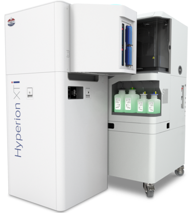
Introducing new groundbreaking capabilities for multiplexed tissue imaging at never-before-seen speeds and automation. The Hyperion™ XTi Imaging System is now available with three imaging modes for rapid and detailed analysis and automated slide loading for walk-away automation.
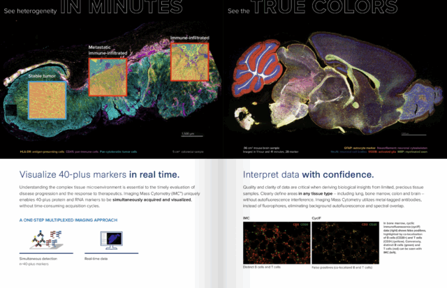
Powered by proven Imaging Mass Cytometry™ (IMC™) technology, the Hyperion XTi Imaging System is a multiplexed tissue imager that uses metal-tagged antibodies, instead of fluorophores, to simultaneously and reliably acquire 40-plus protein and RNA markers, without autofluorescence interference.
Image 40-plus markers on 40 slides in 24 hours.
Expand the possibilities of your research with the Hyperion XTi Imaging System
- Image any tissue type with no autofluorescence interference
- Automated slide loading for walk-away acquisition
- Three acquisition modes to accommodate any type of research
- Batch staining workflow for high-volume studies
- Interpret high-plex data quickly with multiple acquisition modes
Book a virtual demo