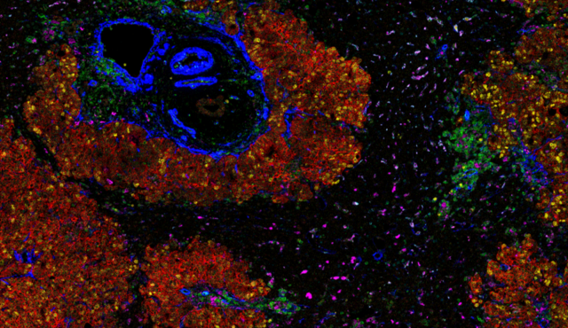
See more in every tissue sample with Spatial Multiomics
Understanding tissue biology requires more than a single data modality. Same-slide omics is a multimodal spatial biology approach that combines complementary molecular, morphological and functional measurements from the same tissue section. By adding high-plex spatial proteomics after RNA, H&E or other protein analysis workflows, researchers can connect gene expression, tissue morphology, molecular composition and protein function while preserving spatial context.
A Flexible Platform for Multimodal Spatial Biology
The Hyperion™ XTi Imaging System enables spatial proteomic analysis using Imaging Mass Cytometry™ (IMC™) following upstream workflows including:
- Spatial transcriptomics
- H&E staining
- MALDI imaging mass spectrometry
- Other morphology-driven tissue imaging approaches
| Technology | Primary measurement | Biological insight | Integration with IMC |
|---|---|---|---|
| Spatial transcriptomics | RNA expression | Gene expression and cellular states | Connect transcriptional programs with spatial protein expression and functional state |
| H&E staining | Tissue morphology | Pathology, tissue architecture and region selection | Add high-plex phenotyping within histologically defined regions |
| MALDI-MSI | Metabolites, lipids and other molecular species | Molecular composition and metabolic heterogeneity | Relate molecular distributions to cell phenotype and tissue organization |
By applying high-plex proteomic analysis after these complementary modalities, researchers can maximize information obtained from valuable tissue samples while preserving spatial context across multiple biological layers.
Spatial transcriptomics and IMC
Connect gene expression to functional biology
This workflow allows researchers to combine transcriptional and functional protein information from the same slide without sacrificing precious tissue samples.
- Validate transcript findings at the protein level
- Improve cell-type identification
- Characterize immune and tumor microenvironments
- Investigate RNA-protein discordance
- Generate richer biomarker discovery datasets
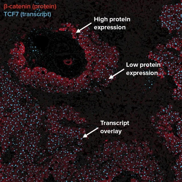 High-plex imaging of protein and RNA on the same slide
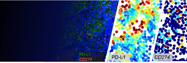
Can RNA and protein be measured on the same tissue section? Absolutely. IMC-based spatial proteomics can follow compatible spatial transcriptomic workflows, enabling RNA and protein analysis from the same tissue section.
Why measure RNA and protein together? Spatial transcriptomics reveals where genes are expressed within a tissue, providing critical insight into cellular states and tissue organization. However, transcript abundance does not always correlate with protein expression. By imaging both, researchers capture complementary layers of cell activity.
Read this application note to learn more about gaining multiomic information-rich data from each tissue slide.
Standard BioTools is proud to announce a new partnership with the Asia-Pacific Spatial Translational Research Alliance (ASTRA), in which Imaging Mass Cytometry™ has been selected as ASTRA’s preferred standard for spatial proteomics.
ASTRA, a consortium of leading cancer researchers across the Asia-Pacific region, is working to build an astounding 5,000 sample pan-cancer spatial atlas aimed at advancing our understanding of oncofetal cell states, therapeutic response and tumor biology. Studies will focus on gathering data from diverse Asia-Pacific populations, filling an important gap in current cancer metrics.
By integrating high-dimensional spatial proteomics into this landmark initiative, researchers will be able to complement spatial transcriptomic insights with quantitative protein-level and cellular phenotyping data, enabling a more comprehensive view of cellular communities and their interactions in cancer development and drug response.
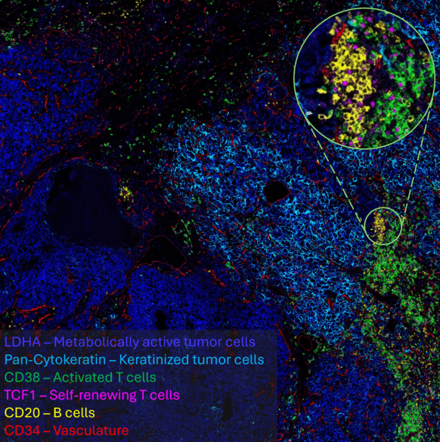
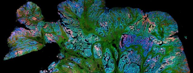
This image was generated using a unique ASTRA 40-marker IMC panel, with the initial antibody selection meeting to images completed within 3 weeks.
H&E staining and IMC
Link tissue morphology with high-plex protein expression
Hematoxylin and eosin (H&E) staining remains the gold standard for tissue morphology assessment. Pathologists rely on H&E to identify tissue architecture, disease features and regions of interest.
A spatial proteomics workflow can be incorporated after H&E imaging, enabling researchers to:
- Preserve conventional pathology workflows
- Select regions of interest based on morphology
- Correlate tissue structure with protein expression
- Compare pathology findings with spatial proteomic data
- Support translational and biomarker studies
This approach bridges traditional histopathology with next-generation spatial biology, generating deeper biological insights from a single tissue section. View this poster highlighting the integration of H&E histology with spatial proteomic profiling.
Connect with a specialist →
MALDI mass spectrometry imaging and IMC
Integrate spatial metabolomics and spatial proteomics
MALDI mass spectrometry imaging (MALDI-MSI) enables spatial mapping of metabolites, lipids and other molecular species throughout tissue sections. When combined with IMC, researchers gain complementary insight into both molecular composition and protein expression.
A MALDI-to-IMC workflow can help researchers:
- Correlate metabolic pathways with protein expression
- Investigate disease-associated molecular networks
- Study tumor metabolism and immune interactions
- Characterize tissue heterogeneity
- Expand biomarker discovery opportunities
Adding IMC after MALDI connects metabolite and lipid distributions with protein expression and cellular phenotypes in the same tissue section, creating a more complete spatial biology workflow. Read this publication combining MSI-based metabolomics and IMC-based immunophenotyping on a single tissue section to reveal metabolic heterogeneity at single-cell resolution.
Connect with a specialist →
How same-slide omics works: IMC offers flexibility to complement other spatial modalities using the same sample
Spatial proteomics can easily be added after transcriptomic imaging and/or an H&E workflow. The workflow directly follows transcriptomic acquisition on the same slide for a critical additional layer of spatial proteomic data. H&E staining can be incorporated between transcriptomic and proteomic workflows, if desired.
Protein markers are stained and acquired simultaneously. This helps minimize tissue degradation prior to acquisition, greatly reduces assay development requirements and helps make this type of multi-omic workflow not only possible, but routine.
Presented by Ankur Sharma at the Garvan Institute of Medical Research, this session applies spatial transcriptomics and protein analysis to oncofetal biology, revealing shared cellular programs between fetal development and cancer that drive proliferation, immune evasion and disease progression. These approaches are already informing a phase 2b liver cancer trial and a growing pan‑cancer spatial atlas, underscoring the importance of integrated RNA–protein analysis for clinical translation and biomarker discovery.
Read the Publication: An Integrated Spatial Multi-Omics Workflow for Sequential RNA and Protein Profiling in FFPE Tumor Tissue
Mainthan Palendira and Ellis Patrick from the University of Sydney present their innovative research combining spatial transcriptomics (Xenium platform) with IMC to study human immune responses in tissues and tumors. This seminar is divided into two parts: the first explores immunology concepts and the technical approach of running both Xenium and IMC on the same tissue section; the second part focuses on data analysis, examining discrepancies between protein and transcript detection at the single-cell level.

Experiment considerations
For more detailed information on this workflow and how to plan your experiment, read the application note Combining Spatial Transcriptomics and Hyperion XTi Workflows for More Comprehensive Spatial Biology, which provides an overview of the application to detect protein targets using IMC analysis after transcript detection from the same slide. With the versatility of IMC technology, the workflow allows any antibodies of interest to be added, or pre-configured and validated IMC panel sets to be used.
Download app noteWhy use Hyperion systems for same-slide omics?
Assay development time: Because IMC technology uses an antibody reagent cocktail, it is simple to mix and match different antibodies without extensive assay validation.
Linear dynamic range and quantitation: Brightfield imaging (DAB) gives at best one order of magnitude (OoM) of dynamic range, while fluorescence gives 2–3 OoM and IMC technology gives 5 OoM. This enables the quantitation of weak signals in the presence of bright signals in the same sample.
Flexibility of tissue acquisition parameters (imaging modes) and a 40-slide autoloader: Capture data from 40 slides in 24 hours, and run your whole study in an automated walk-away run.

How scientists use same-slide omics
Key publications showcasing IMC technology
Additional support: For support information and help with adapting the IMC protocol to your application, talk with one of our Field Applications Scientists here.
Frequently asked questions
Same-slide omics is a multimodal spatial biology approach that enables multiple complementary datasets to be generated from a single tissue section through the sequential analysis of biological features, such as RNA, proteins, morphology or metabolites. By integrating modalities such as spatial transcriptomics, Imaging Mass Cytometry protein imaging, H&E morphology, and other spatial analyses, researchers can gain a more comprehensive view of tissue organization, cellular function and disease biology.
Though mRNA and protein levels are related, they often diverge due to regulatory controls, timing delays and biological noise. A fixed tissue section offers only a snapshot in time, and RNA and protein abundances may not align at that moment. By imaging both, researchers capture complementary layers of cell activity. This dual approach unlocks multidimensional insights into cell states, tissue organization and disease mechanisms. Standard BioTools™ Lab Services support this integrated workflow to help maximize the value of every sample.
Each modality measures a different aspect of biology. Combining them provides a more comprehensive understanding of cell function, tissue organization and disease mechanisms.
For example, combining spatial transcriptomics with high-plex proteomic imaging provides a more complete view of tissue biology than either modality alone. Transcriptomic imaging reveals gene expression activity, while proteomics captures functional state – what the cell is actually doing. Because immune cell phenotypes are typically defined by protein expression, not RNA, cell identity can only be inferred from transcripts but is confirmed through protein detection.
Spatial transcriptomics is a method enabling high-dimensional investigation of gene transcription in a spatial context. By characterizing expression profiles, spatial transcriptomics is important for analyzing transcriptional patterns and regulation as well as identifying cellular neighborhoods and characteristics contributing to disease. Spatial proteomics, on the other hand, is the method of acquiring high-dimensional images of protein expression in a spatial context.
While single-cell transcriptomics (or scRNA-seq) has become a standard in clinical and translational research, the need to preserve intact and viable cells excludes many cell types and destroys any organizational learnings. Spatial transcriptomics addresses these limitations, providing a comprehensive understanding of cell identity and function relative to neighboring cells. Similarly, while regular pathology proteomics (for example, regular DAB IHC) is used to investigate protein expression in tissues, spatial proteomics enables the analysis of 40 or more markers in a single tissue section.
Combining spatial transcriptomics with Imaging Mass Cytometry allows researchers to measure both RNA and protein expression in the same tissue section, helping identify cell states, validate biomarkers and better understand tissue biology.
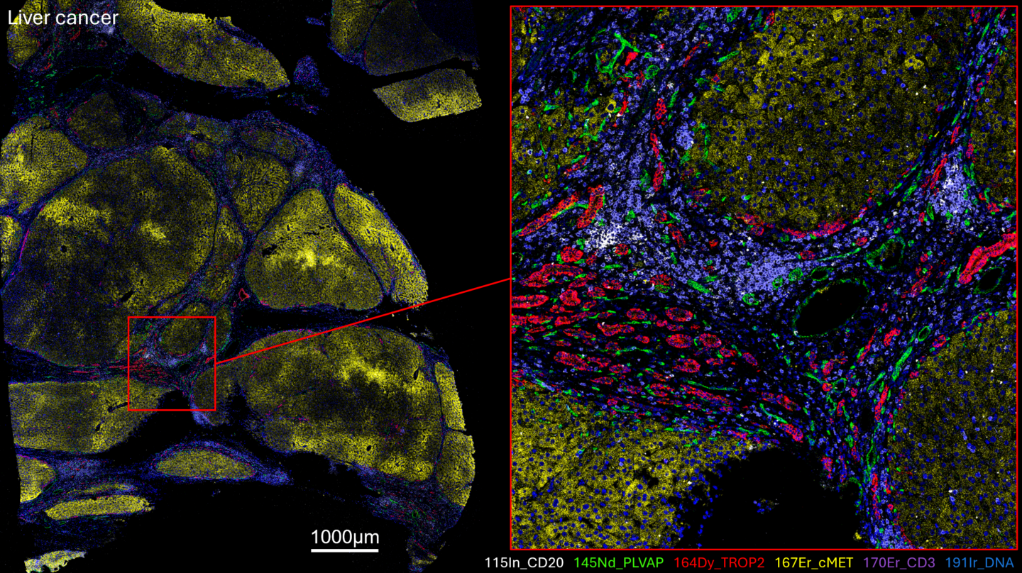
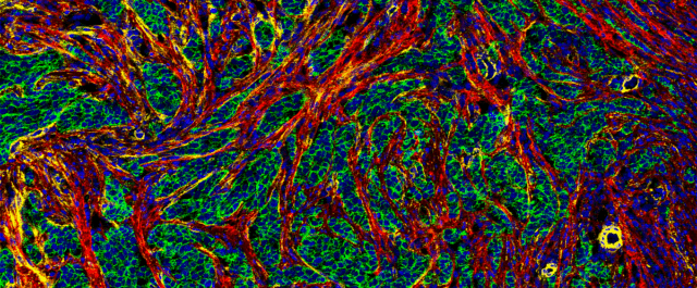
IMC platforms enable the comparison of multiomic data with a choice of up to 45 spatial proteomic markers in the same section. For example, this image of clear cell renal cell carcinoma tissue shows six of 43 markers simultaneously detected in the same multiplexed scan.
The IMC workflow can easily follow other workflows on the same slide to deliver high-quality complementary data. Seamless integration of approaches is enabled by the stability of metal-tags, the simplicity of the IMC workflow and the existence of available integrated data analysis solutions. Cell phenotyping capabilities for tumor and immune components of the tissues remain intact in post-transcript detection slides imaged with IMC platforms. H&E staining can be incorporated between spatial transcriptomics and IMC application to generate data for pathological review of tissue.