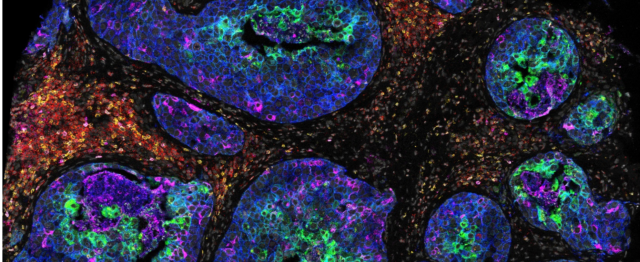
IMC reveals protein biomarkers, resistance mechanism and combination strategies
Antibody-drug conjugates (ADCs) have become one of the fastest-growing areas in oncology drug development. By combining the specificity of monoclonal antibodies with potent cytotoxic payloads, ADCs are designed to deliver tumor-killing agents to cells expressing a selected target. They have delivered transformative clinical benefit in diseases such as HER2-positive breast cancer and are expanding across a growing number of tumor types. Yet despite the promise for precision oncology, ADC response remains variable, as target expression alone often fails to explain why some tumors respond while others exhibit resistance. For pharmaceutical researchers developing ADCs, key questions remain: What mechanisms drive therapeutic response and resistance? How does the tumor microenvironment (TME) influence drug delivery and efficacy? What combination strategies could improve response?
Going beyond RNA: Spatial proteomics adds functional context important for ADC biology
While genomic and transcriptomic approaches provide valuable insights into cancer biology, they do not directly capture protein abundance, activation state, post-translational modifications or cell-cell interactions, all of which are emerging as important influencers in therapeutic response. Imaging Mass Cytometry™ (IMC™) technology helps bridge this gap by providing a functional view of tumor biology, enabling researchers to investigate cellular organization, biomarker distribution, cellular interactions and biological mechanisms.
A recent study published in Cell Reports Medicine demonstrates the value that spatial proteomics can bring to ADC research. Building on a previous spatial transcriptomic analysis of the same cohort, researchers used a 35-marker IMC panel1 to profile more than 912,000 single cells from 47 patients with HER2-positive, hormone receptor-negative breast cancer treated with the HER2-targeted ADC SHR-A1811 in the FASCINATE-N clinical trial. IMC analysis enabled the measurement of protein-level and post-translational features associated with therapeutic response, providing biological insight that could not be directly captured through transcriptomic analysis alone.
Key study finding #1: A post-translational protein IMC readout became central to identifying a response
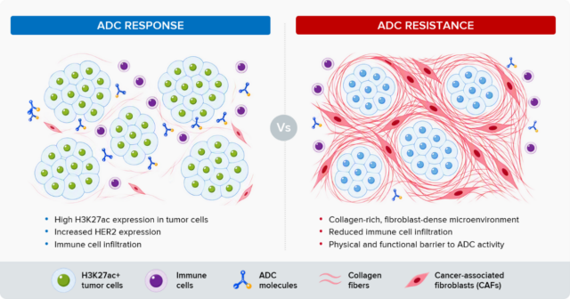
In this study, patients who achieved a pathological complete response had tumors containing higher proportions of H3K27ac-positive epithelial cells. H3K27ac is a post-translational histone modification associated with active transcription, and mechanistic studies show that its enrichment increases HER2 expression, potentially enhancing target availability for anti-HER2 ADCs. Because H3K27ac cannot be measured directly by spatial transcriptomics, this finding highlights the value of spatial proteomics for measuring biologically relevant protein states that RNA-based approaches may miss.
Key study finding #2: Spatial context shapes ADC response
Response is influenced not only by the target but also by the spatial organization of the TME. Using IMC technology to map cellular phenotypes and tissue architecture in situ, researchers found that tumors enriched with collagen-positive fibroblasts were more likely to exhibit resistance. Spatial analysis linked these cells to collagen-rich environments associated with reduced immune infiltration, increased immune suppression and physical barriers to ADC activity. Tumor architecture, immune-cell organization, stromal barriers and protein activity states can all influence whether an ADC reaches its target and delivers its payload effectively.
Key study finding #3: IMC can be used to inform patient stratification and investigate combination strategies
Spatial features derived from IMC data can be incorporated into research frameworks that can help stratify patients more likely to benefit from ADC therapy. The authors introduced an ADC barrier prediction model, which integrates spatial relationships derived from IMC among fibroblasts, endothelial cells and epithelial cells to characterize resistance-associated barriers to ADC response. Lower scores were associated with tissue environments more favorable to ADC activity, while higher scores reflect spatial features associated with resistance.
These same spatial features also supported combination therapy hypotheses designed to improve response. Guided by the IMC findings, the researchers evaluated chidamide to increase H3K27ac levels and losartan to target collagen-rich stromal barriers associated with resistance. In preclinical models, both approaches enhanced ADC activity by increasing HER2 expression or improving immune infiltration within the TME.
From protein biomarker discovery and resistance biology to patient stratification and combination therapy hypotheses, this study illustrates how spatial proteomics can provide functional context that complements transcriptomic analysis and informs ADC development.
Bringing spatial proteomics into routine pathology workflows
A practical question for translational researchers is how these complementary protein-level insights can be incorporated into existing histology-based workflows. While spatial proteomics adds a new dimension to tissue analysis, it does not require abandoning conventional histopathology. In fact, the authors emphasize the value of integrating high-plex spatial proteomics with established histopathology workflows.
“… combining standardized H&E-based TIL evaluation with high-plex spatial proteomic approaches, such as the IMC platform used in this study, provides complementary information and likely represents the future direction for precision immuno-oncology.”
While H&E staining provides critical morphologic context, it does not enable multiplex phenotyping or spatial assessment of cell-cell interactions.
A workflow review: Integrating H&E and IMC on the same FFPE tissue section
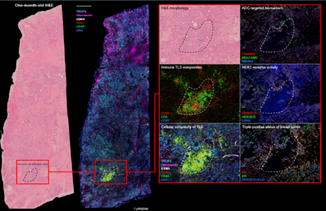
A recent poster highlights an integrated same-slide H&E and IMC workflow for FFPE tissue analysis, demonstrating how conventional histology and high-plex spatial proteomics can be combined on the same tissue section for multiplexed profiling of ADC biomarkers. H&E-stained slides are first imaged to capture tissue morphology and regions of interest before undergoing IMC staining and multiplexed imaging. The resulting IMC data can then be computationally aligned with the original H&E image, enabling integrated analysis of tissue architecture, cellular phenotypes and biomarker expression.
Researchers demonstrated this workflow on both freshly stained and archival H&E slides, including samples stored for approximately one, two and 10 years. By combining conventional histology with high-plex spatial proteomics, researchers mapped ADC-relevant biomarkers including HER2, TROP2, Nectin-4, Claudin 6, CEACAM5, mesothelin, ER and PR within their tissue context. The approach also revealed spatial heterogeneity and microenvironmental features that were not apparent from morphology alone, while simultaneously characterizing tumor, immune, stromal and metabolic features from a single tissue section.
What spatial proteomics could mean for precision oncology
For ADC developers, this broadens the biomarker question from whether the target is expressed to which functional biological states regulate that target and influence its availability to therapy. Spatial proteomics may help researchers understand not only who responds to an ADC, but also why. Researchers at Gustave Roussy have incorporated an antibody that recognizes the ADC payload into IMC assays, allowing them to visualize where ADC-derived drug remains within FFPE tissue. This approach enables direct investigation of ADC distribution, cellular uptake, DNA damage responses and immune activation in spatial context.
While these examples focus on ADC development, the same principles extend to other targeted therapeutic modalities, including small-molecule inhibitors, immune checkpoint therapies, cell therapies and radiopharmaceuticals. As precision oncology continues to evolve, spatial proteomics is becoming an increasingly valuable tool for connecting target expression, protein function, tissue architecture and drug activity to better understand the biological mechanisms that influence therapeutic efficacy.
Resources
- Cai, Y-W. et al. “Spatial proteogenomic profiling uncovers sensitization strategies for antibody-drug conjugate in HER2-positive breast cancer.”Cell Reports Medicine 7 (2026): 102987.
- Natale, D. et al. Integrating Hematoxylin and Eosin Histology with Multiplexed Imaging Mass Cytometry for Spatial Proteomic Profiling of Antibody-Drug Conjugate Targeted Biomarkers (2025).
- Pistilli, B. et al. “Patritumab deruxtecan in HR+HER2− advanced breast cancer: a phase 2 trial.” Nature Medicine 31 (2025): 3,492–3,503.
- Planchard, D. et al. “Efficacy, safety, and biomarker analysis of datopotamab deruxtecan in advanced non-small cell lung cancer: ICARUS-LUNG01 phase 2 study.” Cancer Cell 44 (2026): 1,147–1,159.e6.
- Wijerathna-Yapa, A. et al. “Spatial multiomics approaches for antibody-drug conjugate target discovery.” Cell Press Blue 1 (2026): 100085.
__________________________________________________________
- The 35-marker IMC panel in this study included: tumor and ADC-relevant markers (HER2, HER3, EGFR, pan-cytokeratin and MHC class I); immune markers (CD3, CD4, CD8, CD20, CD56, CD68, CD74, CD11c, CD14, CD206, FoxP3, PD-1, MPO and tryptase); stromal and vascular markers (αSMA, FAP, PDGFRA, PDPN, collagen 1, CD31 and S100A4); and functional protein-state markers (H3K27ac, O-GlcNAc, γH2AX, STING, cGAS, pS6, m6A and Ki-67).