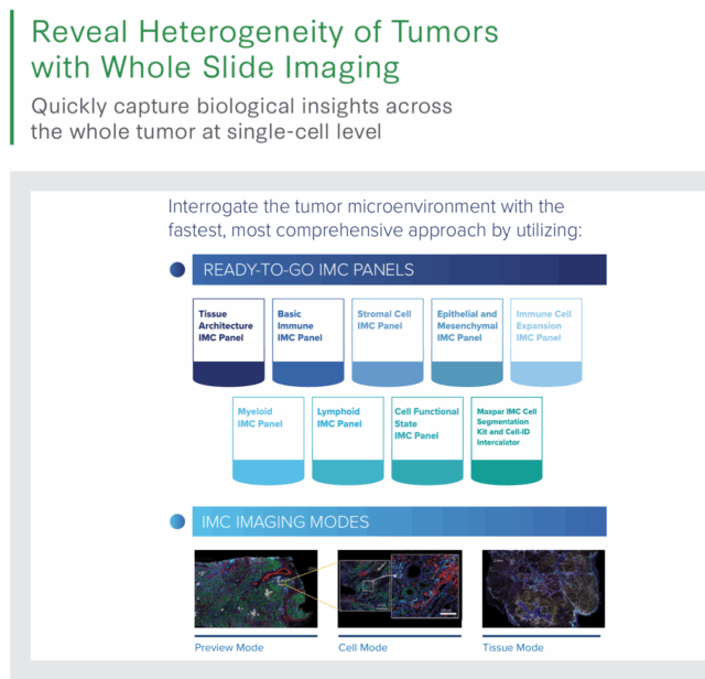
u003ch5u003eu003cstrongu003eVisualize u003c/strongu003ethe immune response. u003cstrongu003eIdentify u003c/strongu003espatial biomarkers. u003cstrongu003eAssess u003c/strongu003edisease progression.u003c/h5u003ernCombine carefully curated IMC panels and cell segmentation markers to study more than 40 targets in a single experiment. Get answers faster with streamlined setup and insights tailored to your research in cancer biology, therapeutic response and disease mechanisms.rnrnu0026nbsp;rnu003cdiv class=u0022container contentu0022u003ernu003cdiv class=u0022py-5u0022 style=u0022display: flex; gap: 1rem; justify-content: center; flex-wrap: nowrap;u0022u003ernrnu003c!u002du002d Card 1 u002du002du003ernu003cdiv class=u0022col-12 col-sm-6 col-lg-3u0022u003ernu003cdiv class=u0022card h-100u0022 style=u0022border-radius: 8px; border: 1px solid #C4C4C4; background: radial-gradient(82.81% 33.43% at 48.05% 105.03%, rgba(28, 101, 0, 0.20) 0%, rgba(255, 255, 255, 0.20) 100%), #FFF; box-shadow: 0px 4px 4px 0px rgba(0, 0, 0, 0.25); padding: 24px;u0022u003ernu003ch3 class=u0022color-secondary-blueu0022u003eu003cstrongu003eReady-to-useu003c/strongu003eu003c/h3u003ernrnu003chr /u003ernrnSpend less time on complicated panel design and optimization with turnkey panels for faster results. rnrnu003c/divu003ernu003c/divu003ernu003c!u002du002d Card 2 u002du002du003ernu003cdiv class=u0022col-12 col-sm-6 col-lg-3u0022u003ernu003cdiv class=u0022card h-100u0022 style=u0022border-radius: 8px; border: 1px solid #C4C4C4; background: radial-gradient(82.81% 33.43% at 48.05% 105.03%, rgba(28, 101, 0, 0.20) 0%, rgba(255, 255, 255, 0.20) 100%), #FFF; box-shadow: 0px 4px 4px 0px rgba(0, 0, 0, 0.25); padding: 24px;u0022u003ernu003ch3 class=u0022color-secondary-blueu0022u003eu003cstrongu003eBroadrncoverageu003c/strongu003eu003c/h3u003ernrnu003chr /u003ernrnComprehensive assessment of cell signaling or metabolic activity targets in a single panel.rnrnu003c/divu003ernu003c/divu003ernu003c!u002du002d Card 3 u002du002du003ernu003cdiv class=u0022col-12 col-sm-6 col-lg-3u0022u003ernu003cdiv class=u0022card h-100u0022 style=u0022border-radius: 8px; border: 1px solid #C4C4C4; background: radial-gradient(82.81% 33.43% at 48.05% 105.03%, rgba(28, 101, 0, 0.20) 0%, rgba(255, 255, 255, 0.20) 100%), #FFF; box-shadow: 0px 4px 4px 0px rgba(0, 0, 0, 0.25); padding: 24px;u0022u003ernu003ch3 class=u0022color-secondary-blueu0022u003eu003cstrongu003eUniquelyrnmodularu003c/strongu003eu003c/h3u003ernrnu003chr /u003ernrnFor use alone or combined with compatible panels. Only with IMC technology.rnrnu003c/divu003ernu003c/divu003ernu003c!u002du002d Card 4 u002du002du003ernu003cdiv class=u0022col-12 col-sm-6 col-lg-3u0022u003ernu003cdiv class=u0022card h-100u0022 style=u0022border-radius: 8px; border: 1px solid #C4C4C4; background: radial-gradient(82.81% 33.43% at 48.05% 105.03%, rgba(28, 101, 0, 0.20) 0%, rgba(255, 255, 255, 0.20) 100%), #FFF; box-shadow: 0px 4px 4px 0px rgba(0, 0, 0, 0.25); padding: 24px;u0022u003ernu003ch3 class=u0022color-secondary-blueu0022u003eu003cstrongu003eDynamicrnrangeu003c/strongu003eu003c/h3u003ernrnu003chr /u003ernrnDetect high- and low-expressing markers with Hyperion™ systems. rnrnu003c/divu003ernu003c/divu003ernu003c/divu003ernu003c/divu003ernu0026nbsp;
u0026nbsp;rnu003ch3 style=u0022text-align: center;u0022u003eIMC panelsu003c/h3u003e
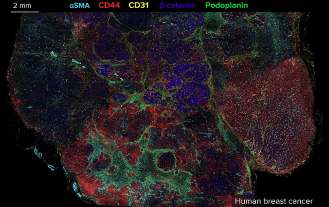
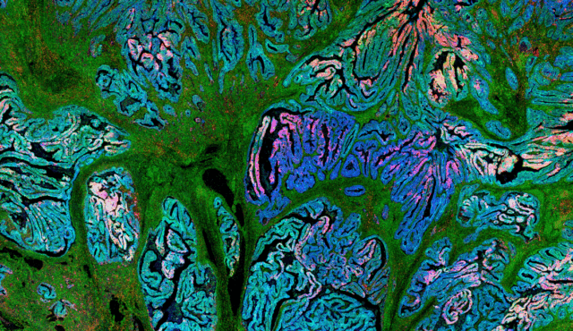
u003ch3 data-gtm-vis-first-on-screen11876973_193=u00224997u0022 data-gtm-vis-total-visible-time11876973_193=u00221000u0022 data-gtm-vis-has-fired11876973_193=u00221u0022u003eu003c/h3u003ernu003ch3 data-gtm-vis-first-on-screen11876973_193=u00224997u0022 data-gtm-vis-total-visible-time11876973_193=u00221000u0022 data-gtm-vis-has-fired11876973_193=u00221u0022u003eu003cstrongu003eHuman immuno-oncologyu003c/strongu003eu003c/h3u003ernThe Human Immuno-Oncology IMC Panel, 31 Antibodies combines overall tissue architecture, cell interaction and single-cell phenotypic and functional markers in the single largest Imaging Mass Cytometry panel currently available.rnrnu003ca class=u0022secondary-btnu0022 href=u0022https://dam.standardbio.com/asset/f355fb41-b397-4afc-b222-f265bac78858/Human-Immuno-Oncology-IMC-Panels-Flyer-FLDM-01335-Rev-05.pdfu0022 target=u0022_blanku0022 rel=u0022noopeneru0022u003eSee panel applications and available markersu003c/au003ernrnu003ca class=u0022secondary-btnu0022 href=u0022https://dam.somalogic.com/m/13897f889df4fe8a/original/Reveal-Heterogeneity-of-Tumors-with-Whole-Slide-Imaging-Application-Note-082025-FLDM-01344-Rev-02.pdfu0022 target=u0022_blanku0022 rel=u0022noopeneru0022u003eRead the application noteu003c/au003ernu003ch6u003eComprehensive panel includes: PanCK, CD31, Collagen 1, Fibronectin, FAP, Podoplanin, αSMA, CD44, CD45, CD3, CD20, CD68, CD4, CD8, CD45RO, CD57, CD66b, HLA-DR, CD163, CD14, CD11b, CD11c, Granzyme B, PD-L1, PD-1, FoxP3, Ki-67, E-cadherin, β-catenin, EpCAM, Vimentinu003c/h6u003e
u003cimg class=u0022alignnone size-ra_small wp-image-7579u0022 src=u0022https://standardbio.com/wp-content/uploads/2026/01/Human-IO-panel-image-640×404.pngu0022 alt=u0022u0022 width=u0022640u0022 height=u0022404u0022 /u003e
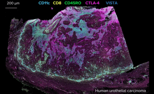
u003ch3 style=u0022text-align: left;u0022 data-gtm-vis-first-on-screen11876973_193=u00224997u0022 data-gtm-vis-total-visible-time11876973_193=u00221000u0022 data-gtm-vis-has-fired11876973_193=u00221u0022u003eu003c/h3u003ernu003ch3 style=u0022text-align: left;u0022 data-gtm-vis-first-on-screen11876973_193=u00225424u0022 data-gtm-vis-total-visible-time11876973_193=u00221000u0022 data-gtm-vis-has-fired11876973_193=u00221u0022u003eu003cstrongu003eT cell exhaustionu003c/strongu003eu003c/h3u003ernu003cp style=u0022text-align: left;u0022u003eAssess immune dysfunction in chronic viral infections and cancer and help guide development of effective immunotherapies with the Human T Cell Exhaustion IMC Panel, 5 Antibodies.u003c/pu003ernu003cp style=u0022text-align: left;u0022u003eu003ca class=u0022secondary-btnu0022 href=u0022https://dam.standardbio.com/asset/f355fb41-b397-4afc-b222-f265bac78858/Human-Immuno-Oncology-IMC-Panels-Flyer-FLDM-01335-Rev-05.pdfu0022u003eSee panel applications and available markersu003c/au003eu003c/pu003ernu003cp style=u0022text-align: left;u0022u003eu003ca class=u0022secondary-btnu0022 href=u0022https://dam.somalogic.com/m/248b32c25caff2da/original/CyTOF-Summit-2025-Human-IO-T-cell-Exhaustion.pdfu0022 target=u0022_blanku0022 rel=u0022noopeneru0022u003eSee panel datau003c/au003eu003c/pu003ernrnu003ch6u003eT cell-specific panel includes: TIM-3, IDO, LAG-3, OX40, CTLA-4u003c/h6u003e
u003cimg class=u0022alignnone size-ra_small wp-image-7581u0022 src=u0022https://standardbio.com/wp-content/uploads/2026/01/T-cell-exhaustion-panel-image-640×391.pngu0022 alt=u0022u0022 width=u0022640u0022 height=u0022391u0022 /u003e
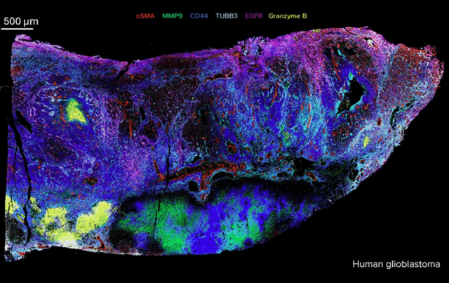
u003ch3 data-gtm-vis-first-on-screen11876973_193=u00224997u0022 data-gtm-vis-total-visible-time11876973_193=u00221000u0022 data-gtm-vis-has-fired11876973_193=u00221u0022u003eu003c/h3u003ernu003ch3 data-gtm-vis-first-on-screen11876973_193=u0022585300u0022 data-gtm-vis-total-visible-time11876973_193=u00221000u0022 data-gtm-vis-has-fired11876973_193=u00221u0022u003eu003cstrongu003eHuman neuro-oncologyu003c/strongu003eu003c/h3u003ernTarget neuronal-specific markers in human gliomas with the Glioblastoma IMC Panel, 5 Antibodies. Reveal spatial complexity in heterogeneous brain tissue without tissue degradation and autofluorescence artifacts usually observed in brain tissue.rnrnu003ca class=u0022secondary-btnu0022 href=u0022https://dam.somalogic.com/m/48291ce339ca3365/original/Whole-Slide-Imaging-Modes-and-Curated-Antibody-Panels-for-Imaging-Mass-Cytometry-Approach-Reveal-Extensive-Spatial-Heterogeneity-of-Human-Glioblastomas-Horizontal-Poster-072025.pdfu0022 target=u0022_blanku0022 rel=u0022noopeneru0022u003eSee example data and available markersu003c/au003ernrnu003ca class=u0022secondary-btnu0022 href=u0022https://fluidigm.my.salesforce.com/sfc/p/700000009DAw/a/QQ000001VcGX/dp1BkPzzjzLtvtFHFZiUjmy_rgp5fhH6MMo0fz1Elwwu0022 target=u0022_blanku0022 rel=u0022noopeneru0022u003eView panel applicationsu003c/au003ernu003ch6u003eGlioblastoma-specific panel includes: EGFR, Vimentin, SOX2, Nestin, MMP9u003c/h6u003e
u003cimg class=u0022alignnone size-ra_small wp-image-7580u0022 src=u0022https://standardbio.com/wp-content/uploads/2026/01/Human-Neuro-panel-image-640×403.pngu0022 alt=u0022u0022 width=u0022640u0022 height=u0022403u0022 /u003e
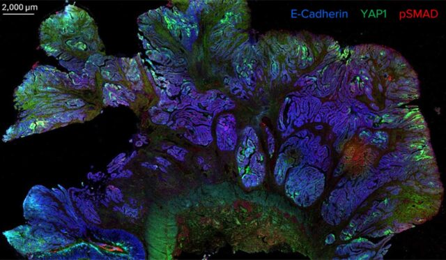
u003ch3 data-gtm-vis-first-on-screen11876973_193=u00224997u0022 data-gtm-vis-total-visible-time11876973_193=u00221000u0022 data-gtm-vis-has-fired11876973_193=u00221u0022u003eu003c/h3u003ernu003ch3 data-gtm-vis-first-on-screen11876973_193=u0022585300u0022 data-gtm-vis-total-visible-time11876973_193=u00221000u0022 data-gtm-vis-has-fired11876973_193=u00221u0022u003eu003cstrongu003eCell signalingu003c/strongu003eu003c/h3u003ernCover multiple signaling pathways in a single panel with the Human Cell Signaling Pathway IMC Panel, 7 Antibodies. Identify pathways regulating key cellular processes such as proliferation, differentiation, survival and tumorigenesis. rnrnu003ca class=u0022primary-btnu0022 href=u0022https://dam.standardbio.com/asset/40c756d9-5861-450a-a4f1-b2c7a0c6ddca/Understanding-Cell-Signaling-Dynamics-in-Cancer-with-Spatial-Proteomics-App-Note-082025-LAB-00069-Rev-1.pdfu0022 target=u0022_blanku0022 rel=u0022noopeneru0022u003eRead the application note for product details and available markersu003c/au003ernrnu003ca href=u0022https://dam.standardbio.com/asset/a718b777-eec4-41bd-b2e7-c8dd8fa417da/Spatial-Proteomics-Profiling-of-Metabolic-and-Cell-Signaling-Heterogeneity-in-Breast-Cancer-Tumor-Poster.pdfu0022u003eSee example datau003c/au003ernu003ch6u003eCell signaling panel includes: EGFR, pSMAD, GSK3β, p-tyrosine, YAP1, pERK 1/2, PTENu003c/h6u003e
u003cimg class=u0022alignnone size-ra_small wp-image-7578u0022 src=u0022https://standardbio.com/wp-content/uploads/2026/01/cellsignalingtissue-cropped-full-640×373.jpgu0022 alt=u0022u0022 width=u0022640u0022 height=u0022373u0022 /u003e
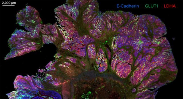
u003ch3 data-gtm-vis-first-on-screen11876973_193=u00224997u0022 data-gtm-vis-total-visible-time11876973_193=u00221000u0022 data-gtm-vis-has-fired11876973_193=u00221u0022u003eu003c/h3u003ernu003ch3 data-gtm-vis-first-on-screen11876973_193=u0022585300u0022 data-gtm-vis-total-visible-time11876973_193=u00221000u0022 data-gtm-vis-has-fired11876973_193=u00221u0022u003eu003cstrongu003eCell metabolismu003c/strongu003eu003c/h3u003ernThe Human Cell Metabolism IMC Panel, 7 Antibodies enables assessment of metabolic activity that plays a critical role in cellular energy production, metabolic adaptation and disease progression, particularly in cancer, immune-mediated dysfunction and metabolic disorders.rnrnu003ca class=u0022primary-btnu0022 href=u0022https://dam.standardbio.com/asset/b6689604-12d9-4030-80a6-0e8060c1cc7d/Unraveling-Metabolic-Reprogramming-in-Cancer-with-Spatial-Proteomics-App-Note-082025-LAB-00068-Rev-2.pdfu0022 target=u0022_blanku0022 rel=u0022noopeneru0022u003eRead the application note for product details and available markersu003c/au003ernrnu003ca href=u0022https://dam.standardbio.com/asset/a718b777-eec4-41bd-b2e7-c8dd8fa417da/Spatial-Proteomics-Profiling-of-Metabolic-and-Cell-Signaling-Heterogeneity-in-Breast-Cancer-Tumor-Poster.pdfu0022u003eSee example datau003c/au003ernu003ch6u003eCell metabolism panel includes: GLUT-1, Citrate synthase, ATP5A, CPT1A, LDHA, p4E-BP1, pS6u003c/h6u003e
u003cimg class=u0022alignnone size-ra_small wp-image-7577u0022 src=u0022https://standardbio.com/wp-content/uploads/2026/01/cellmetabolismtissue-cropped-full-640×349.jpgu0022 alt=u0022u0022 width=u0022640u0022 height=u0022349u0022 /u003ernrnFor detailed information on specific panels, visit the u003ca href=u0022https://store.standardbio.com/px-panel-and-labeling-kits/u0022u003ecatalogu003c/au003e
u003ch4 style=u0022color: #28366a !important; text-align: center;u0022u003eu003cbu003eBuilding a comprehensive panel has never been this easyu003c/bu003eu003c/h4u003ernu003cp style=u0022text-align: center;u0022u003eTo see how all of these panels can fit together, see the u003ca href=u0022https://dam.standardbio.com/asset/da34839b-d89d-4744-abe2-0df513bd3b54/Imaging-Mass-Cytometry-Modular-Panel-Compatibility-Guide-052025-LAB-00055-REV-2.pdfu0022u003eIMC panel compatibility guideu003c/au003e or test out the compatibility of a subset of IMC panels using the interactive builder below.u003c/pu003ernu003cp style=u0022text-align: center;u0022u003eQuickly understand the tissue environment with modular IMC panels that make study design simple, fast and highly flexible. Tailor marker selection to your specific biological pathway or research question by choosing from a suite of 14 carefully curated IMC panels.u003c/pu003e
u003ch4 style=u0022text-align: center;u0022u003eApplicationsu003c/h4u003e

Let us do the work for you.
Did you know Standard BioTools has an in-house service lab? Send us your samples and we will send you the data.rnrnu003ca class=u0022btnu0022 href=u0022https://standardbio.com/products-services/lab-services/u0022u003eLearn more >u003c/au003e
u003ch4 style=u0022text-align: center;u0022u003eResourcesu003c/h4u003e
GET MORE INFORMATION
Interested in Standard BioTools™ Imaging Mass Cytometry products or need a quote? Contact our sales team for product orders, quotes or other inquiries.