

| Key Features | Description |
|---|---|
| High-parameter analysis | Detect 40-plus markers simultaneously in any tissue type without autofluorescence interference |
| Protein and RNA co-detection | Correlate spatial protein expression with transcriptional signatures in the same tissue section |
| Wide dynamic range | Detect both high- and low-abundance targets in a single experiment |
| Ready-to-go panels | Choose from 20-plus pre-optimized, application-specific panels for rapid project startup |
| Custom panels | Select from 800-plus pre-labeled antibodies; custom conjugation is also available |
Services workflow
We support you from sample collection to data delivery
- Section tissue or tissue microarray onto a slide
- Stain slides with one antibody cocktail from Standard BioTools
- Ship FFPE slides at room temperature to our Services Lab
- Multiplexed acquisition of selected regions of interest or entire tissue using IMC technology
- Receive data, including spatial data analytics
Compatible sample types
- Human – Fixed or frozen samples
- Mouse – Fixed or frozen samples
- Whole tissue sections, tissue microarrays (TMAs)
- Tissue can be pre-stained by you prior to shipping or stained by us
Or go back to our Omics Services page.
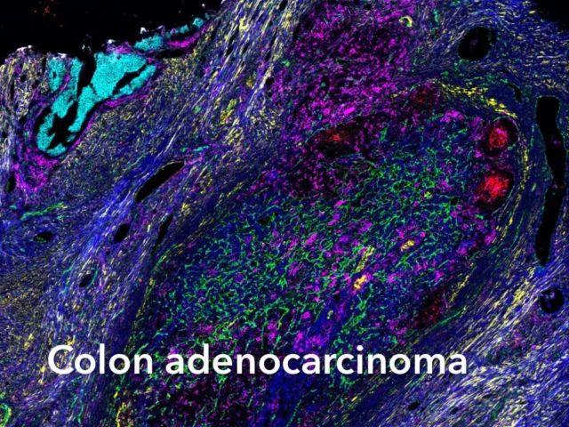
High-resolution IMC images
These figures illustrate the detailed tissue imaging capabilities of the Hyperion™ Imaging System, revealing spatial biology with exceptional clarity.


For Research Use Only. Not for use in diagnostic procedures. Patent and License Information: www.standardbio.com/legal/notices. Trademarks: www.standardbio.com/legal/trademarks. Any other trademarks are the sole property of their respective owners. ©2025 Standard BioTools Inc. All rights reserved.